Main > GMT_cranioplasty
Dr Graham Treece, Department of Engineering
ENGINEERING TRIPOS PART IIA
Module 3G4 - Medical Imaging and 3D Computer Graphics
Titanium cranioplasty
In this work, medical imaging, computer graphics, surface interpolation and computer-controlled milling are used to create titanium cranial implants at Christchurch Hospital, New Zealand.
Researchers: Jonathan Carr, Richard Fright and Rick Beatson, now with Applied Research Associates NZ Ltd .
|
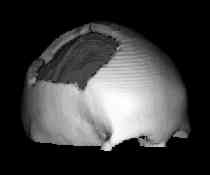
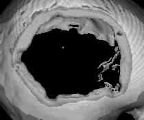
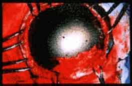
Ray-tracing is used to depict bone surfaces within a stack of CT data slices.
|
|
user graphically identifies a defect in the skull by highlighting the sound bone surrounding the defect
|
|
|
|
|
Radial basis function (RBF) approximation is used to fit a surface to the incomplete depth-map corresponding to the rendered view of the defect. The surface of the skull is smoothly interpolated across the defect. The thin-plate spline basis is chosen in this application.
|
|
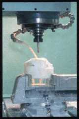
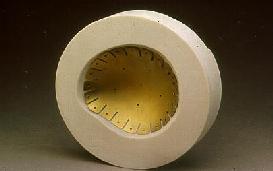
A computer numerical controlled (CNC) mill produces a model of the defect and a mold in the shape of the interpolated surface.
|
|
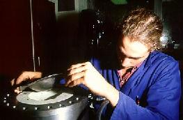
Flat titanium plate is pressed into the mold in a hydraulic press.
|
|
|
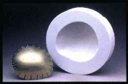
The finished annodised plate fits the mold.
|
|
The perimeter holes in the plate are for the mounting screws and the central holes allow fluid to circulate. Perforations give the plate a limited amount of adjustment in theatre.
|
|
|
The plate is fixed in place with titanium screws.
|
Author: Jonathan Carr.