Main > GMT_4YP_20_3
Dr Graham Treece, Department of Engineering
F-GMT11-3: Assessing trabecular bone over the whole skeleton
 |  |  |
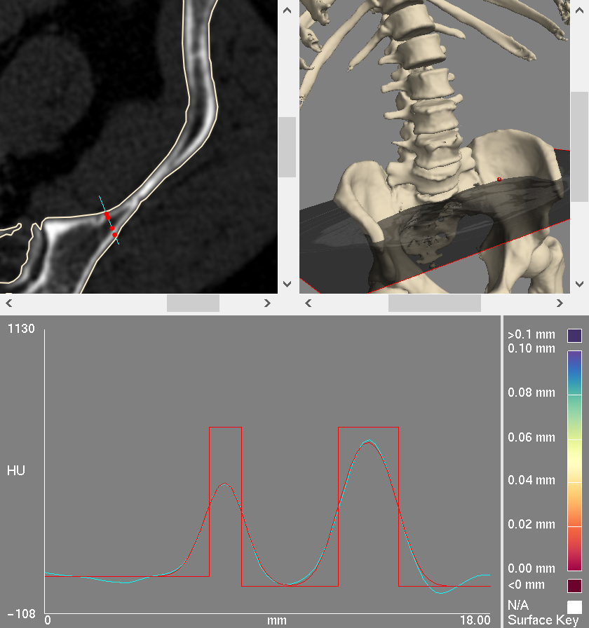
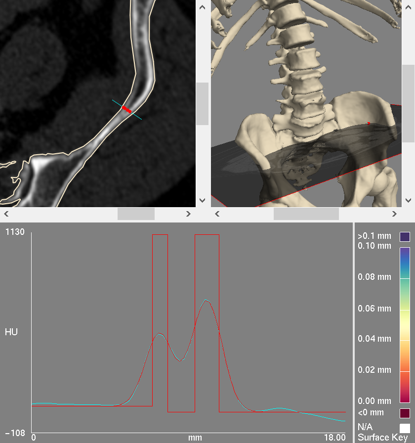
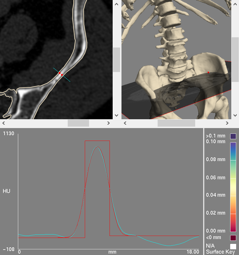
| Here are some cortical (the outer bone surface) bone measurements of a part of the pelvis. These include cortical thickness (the width of each red 'top-hat') and also trabecular (inner bone) density (the height of the red line between the cortices). Such measurements can be very useful for revealing various bone diseases. | In some parts of the skeleton, the cortices are very close together, and it is difficult to measure the trabecular density: how do we know how accurate this measurement is or whether it is possible at all? | In other areas there is only one cortex, and currently the measurements wrongly report soft tissue density as trabecular density. This project aims to produce a reliable trabecular density measurement over the whole skeleton. |
There are a variety of bone diseases which affect many people, and we have recently been involved in research which has the potential to contribute significantly to the accurate detection of small areas of diseased bone. The advances have come from a much more precise (and hence much more sensitive) measurement of the bone cortex (the denser layer surrounding the less dense bone in the middle), and the surrounding trabecular (inner bone) density, collectively known as cortical bone mapping (CBM). CBM has also been used to further our understanding of fracture, underpin models of bone used in mechanical analysis, and is even being used by paleo-anthropologists who are interested in the properties of very old bones.
Most of the work to date has concentrated on measuring the thickness of the cortex, which is expected to be a single 'layer' of bone, but the trabecular density just within the cortex is just as significant. In many places bone has more than one layer: for instance in the pelvis and skull there are two layers: an inner and an outer table, which in some places join together to become just one. In these areas it is not clear whether our measurements relate to one or both tables. The measurement of trabecular density may be missing, or very inaccurate, or wrongly derived from the surrounding soft tissue. The aim of this project is to try to make robust measurements of trabecular bone, by integrating the location of the outer surfaces into the CBM technique. This is an interesting problem in computational geometry, data deconvolution and surface-based visualisation.
This is an algorithmic development / computational geometry / software project, so experience of writing software is essential, though the development could also be done using Matlab.
Click here for other medical imaging projects offered by Graham Treece.


